April 2022

Plastic Bodies
Preservation of the dead is deeply rooted in human nature, from Egyptian mummies to bog bodies to the carefully preserved loved ones we seal away in caskets. While Western culture often separates the living from the dead, there is a traveling, world-renown exhibit with real human bodies on full display: The Body Worlds Exhibit. If you haven’t been to it, you’ve probably heard of it. If you haven’t heard of it, you came to the right place.
There are preserved human bodies, with skin and fat removed, arranged in elaborate and sometimes playful positions. Each specimen is arranged to display a particular feature of human anatomy. Some of the models feature a particular organ or organ system. There is a human form in which only the vascular system is visible. Picture an athlete mid-jump, with quadriceps muscles visible and exploding with power. Imagine seeing every small muscle connecting ribs to one another as a torso twists. A human heart. A uterus. Each tendon in the hand.
We carry our bodies around every day, mostly oblivious to the incredible symphony of structure and function that supports every movement, every breath. Covered in skin, we miss most of the nuance unless something goes wrong. The Body Worlds Exhibit, and the plastination method developed by Gunther von Hagens that makes it possible, create a window into our anatomy. It is an intimate look at what lies beneath.
In 1977, Gunther von Hagens developed the process of plastination. It preserves organic tissue and keeps it from decaying or smelling by converting it to plastic.
First, it’s important to understand that the human bodies this process is performed on are willing donors who, prior to death, agreed to being preserved and displayed. With the potential to become a teaching tool or a component of the Body Worlds exhibit, each donor is informed of the process and the potential uses of his or her body, and often signed up to be donors after visiting the Body World’s exhibit themselves. They chose this process specifically, and were not organ donors who ended up in a museum. Now that the body has been ethically sourced, it has a few steps to take before it’s presentable:
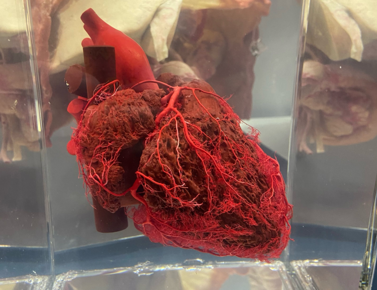
1. The first step is fixation. Often, this is achieved by formaldehyde infusion to kill bacteria or living tissue and to prevent further decay. If a particular vascular system needs to be emphasized, such as in the heart, latex is injected into arteries so they retain their shape and appear lifelike. Fixation typically doesn’t take very long, about 3-5 hours.
2. Dissection to remove skin, connective tissue, and fat from the bodies is necessary in order to prepare the specimen for display of a particular region or organ system. This can take anywhere from 500-1000 hours of skilled dissection. I’ve spent many hours of my own dissecting coronary arterioles and I have immense respect for those who do this for a living.
3. The next step is an acetone bath to remove any remaining fat and water, or to defat and dehydrate. Acetone is the active chemical in nail polish remover, and because it is non-polar molecule, it can dissolve other non-polar molecules, such as fat molecules. At -25 °C, the body is soaked in acetone, replacing the water in each cell with acetone. It mixes incredibly well with water but evaporates much more easily than does water, which come into play in the next step.
4. After all water has been replaced with acetone, the body undergoes forced impregnation of a liquid polymer. Technicians create a vacuum around the body, and the high vapor pressure of acetone causes it to evaporate out of the cells. The liquid polymer has a much lower vapor pressure and does not evaporate but instead, as the acetone evaporates, the polymer is pulled into each cell to plasticize it. This takes 2-5 weeks for a human body. When a 400- pound blue whale heart needed to be preserved, the process took nearly 6 months.
5. The body is then ready for positioning. By this point in the process, the body is quite literally plastic, both in content and pliability, so it is easily manipulated. This is where anatomical knowledge and an artistic eye are essential to pin, clamp, and position each body part where it should be, or in the best position to display a certain system.
6. Finally, the body is cured, or hardened. If silicon oil was the medium that was forced into cells, it has to be exposed to a higher heat in order to harden and become silicon plastic. This final step in the process ensures that nothing decays or decomposes, and that the specimen is durable and long-lasting.
These specimen are incredible teaching tools. There was a booth with many of these anatomical models at the conference I attended last week, and it was amazing to see real human bodies opened up. There were a few that stood out to me. One was a vascular preparation of the heart! It maintained all of the blood vessels with minimal muscle tissue, and it was incredible to see just how dense the vascular system is in the left ventricle, the one responsible for pumping blood to the rest of the body.
A second was a chest/torso with the ribs separated at the front, and the organs pulled forward and out so that the inside of the chest wall was visible, as well as the spine and major nerves. The chest cavity is beautiful and it always surprises me just how many organs can fit inside such a small space.
Finally, there was a hand preparation that emphasized the tendons and ligaments. They were pulled and emphasized so that each layer stood out. Hands get take for granted because we see them so often. Having a human hand, with all its intricate muscle and tendon arrangements, is an incredible teaching tool in addition to being beautiful.
You might not have woken up this morning expecting to learn about plastination, but now you know more and hopefully it’s one of those tidbits that stays with you, the kind you bring up at parties because you can’t stop thinking about how cool it is.